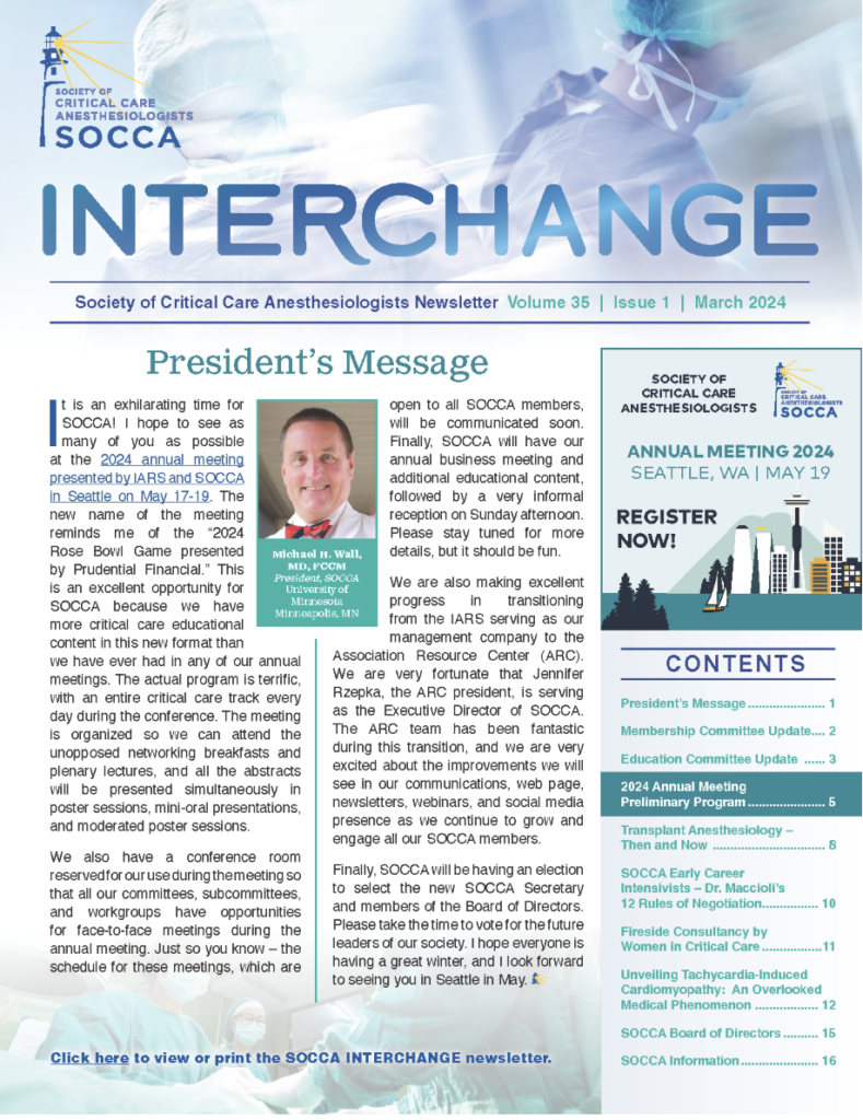
‘Tis the (Flu) Season – Influenza Infection in Critically Ill Patients
As influenza season begins in the United States, we should reflect on the nature of the virus, pathological presentation and treatment considerations. Multiple types of influenza exist, with type A accounting for the most severe manifestations in humans and responsible for the major pandemic outbreaks, notably in 1918, 1957, 1968, and 2009. Types B and C also infect humans but result in a more typical seasonal infection. Type A Influenza is further subtyped based upon surface antigen expression, using the H-N- nomenclature, to describe hemagluttinin and neuraminidase expression. Some subtypes have been found to be notably more pathogenic, such as H1N1, the cause of the “Spanish flu” outbreak in 1918 resulting in an estimated 50 million deaths worldwide. While most strains of the Influenza virus disproportionately affect the young and the old, H1N1 subtypes appear to impact populations more broadly, and they may be more pathologically virulent. Since the 2009 “Swine flu” pandemic, the predominant type A viral subtype has been H1N1.
Regional climate plays a role in viral shedding and influenza infection, with outbreaks in the Southern hemisphere occurring during the winter months (June-August) and then spreading to the Northern hemisphere several months later during the winter months there (January-March of the following year). The nature of the subtropical climates, specifically in southeast Asia, allow for human infection nearly year-round, and birds in these regions may act as a viral reservoir of type A influenza, promoting future yearly worldwide outbreaks. Antigenic shift within strains is partly responsible for pandemic emergence from seasonal strains, and animals may act as a reservoir for these changes. While certain populations will always be susceptible to complications of influenza infection, pandemic strains are often more antigenically novel to humans, and thus result in increased infection numbers and pathogenicity, frequently associated with higher morbidity and mortality.
The 1918 influenza outbreak was notable for several reasons. A pandemic strain emerged into a world developing closer quarters. Military requirements necessitated huge numbers of people in smaller spaces, while urban density was increasing, and sterility was still a novel concept. In addition, a profoundly pathogenic response was provoked by this particular strain of H1N1 influenza, resulting in rampant activation of the native immune response. It was likely that this strain had not been seen in the decades immediately prior, and thus, no limited immunity would have been conferred to the populace. Immunologic response would be expected to be more severe, and it was. Approximately 50 million people died as a result of the 1918 epidemic, likely an underestimation. Successive pandemics have been less severe, relatively, although many factors contribute to that, including improvements in living conditions, health care and treatment options.
The patient with influenza may present differently, depending on age, underlying health status, and timeline of infection. Healthy, young adult patients may take longer to present for medical care and subsequently may be further delayed for aggressive treatment, due to their resilience and compensatory ability. As a result, their illness may be more advanced when admitted. The very young, very old and infirm patients will be more likely to seek early medical care and are much more likely to be treated aggressively.
Several rapid diagnostic influenza tests are currently in use. While some variability exists within tests, nearly all require nasopharyngeal swab sampling and report presence of type A and B viral antigens within 10-30 minutes. The typical rapid influenza diagnostic tests (RIDT) carry high specificity but moderate sensitivity when compared to the gold standard of PCR, potentially allowing for a higher false-negative rate. Digital immunoassay (DIA) and rapid nucleic acid amplification tests (NAAT) allow for higher sensitivity but may introduce increased cost and more technical skill to read the results. Later diagnosis of influenza infection in hospitalized critically ill patients is associated with increases in mortality and secondary complications. One of the most important diagnostic tools in these patients is the clinical index of suspicion, and with the high specificity of the rapid diagnostic tests, it may be reasonable to test more hospitalized patients admitted with respiratory symptoms, whereas outpatients may actually be tested less frequently, depending on timeline of illness.
While the epidemiological assault from the influenza virus is seasonal and predictable, the individual response may not be as easily estimated. As antigenic shift allows for relatively novel subtype presentation, strains that evade immune memory may provoke a worsened host response, resulting in the characteristic cytokine storm and inflammatory cascade that is responsible for much of the systemic damage in severe influenza, as well as promotion of secondary complications. Viral sepsis may be challenging to diagnose and treat, as much of the care is supportive. Immunomodulatory therapies are likely to provides the next weapon against septic viral infections, including influenza, but these are currently experimental. Prompt diagnosis and appropriate critical care management are the mainstay of therapy at this time.
Neuraminidase inhibitors, such as oseltamivir, and the recently approved endonuclease inhibitor baloxovir have been shown to reduce the duration of symptoms in patients with influenza diagnosed within 24-48 hours. Much of the data regarding neuraminidase inhibitor use in critically ill patients is from the most recent 2009 pandemic, the H1N1 “Swine flu”. Neuraminidase inhibitor use has been associated with mortality reduction in critically ill patients with influenza and should be considered in any critically ill patient with influenza, as survival benefit appears to be increased across the board. Higher-dose and extended therapy may be reasonable, especially in patients who remain critically ill or those with progressive symptoms on standard treatment, but data is lacking to strongly support positive impact on survival or ICU-free days. Baloxovir has not been thoroughly evaluated in a critically ill patient population. Older M2 inhibitors (amantadine) are no longer recommended, given the extremely high treatment resistance observed.
Alternative therapies, such as administration of plasma from donors with influenza exposure and intravenous immunoglobulins, show promise in reducing the inflammatory burden and may improve survival, but are likely to be reserved for the sickest patients. Corticosteroids and macrolide compounds such as clarithromycin and rapamycin have defined anti-inflammatory benefits, but their use is not routinely recommended in severe influenza infection, as no obvious benefit has been delineated, and outcomes may actually be worse. Isolation of critically ill patients with influenza should be mandated, and non-clinical exposures limited, given the high risk of transmission. The duration of isolation is institution-specific, but influenza virus has been found in tracheal isolates of intubation patients >21 days after admission.
Bacterial superinfection is commonly associated with severe influenza infection, and may be the presenting complaint in younger patients, as it often tends to develop well after the initial viral infection. Epithelial injury due to viral activity and the associated host response results in a nidus for infection, and community-acquired organisms, such as staphylococcus and streptococcus species, should be suspected. Therapy with a third-generation cephalosporin should be started in patients with post-influenza pneumonia, with or without a fluoroquinolone depending on suspicion. Broad-spectrum antibiotic therapy should always be a consideration in the critically ill patient with infection and is a reasonable approach in the patient with influenza until bacterial superinfection can be ruled out or appropriately speciated.
Epithelial damage, the host response, and bacterial superinfection can all lead to the development pf severe lung injury and ARDS. Management of these patients is fairly consistent with management of ARDS in the general population. Neutrophil migration and endothelial injury promote parenchymal congestion and respiratory failure, either during the initial viral insult or following secondary infection, and strict attention should be given to maintaining low tidal volume ventilation and reduced plateau pressure as able, as outlined in lung-protective ventilation guidelines.
Other approaches to management of severe respiratory failure, such as use of neuromuscular blockade, prone positioning, pulmonary vasodilators and alternative modes of ventilation, may be considered based on the clinical course and institutional abilities. In patients with refractory respiratory failure, ECMO can be considered. Although it can be difficult to prognosticate timeline of the influenza patient with severe respiratory failure, survival is significantly better with earlier application; however, this should be balanced with risks when stratifying patients for selection, and not all patients will be appropriate candidates. Early referral to an ECMO consultation service or ECMO center may help clarify opportunities and expectations in the critically ill patient with influenza.
In closing, the critically ill patient with influenza has a high risk of morbidity and mortality, influenced in part by underlying health status, severity of pandemic outbreak, bacterial superinfection, and host response to infection. Early recognition and aggressive treatment is imperative in this patient population.
Author

Categories
- Announcements
- Annual Meeting
- Award
- Book & Media Reviews
- Call for Articles
- Case Reports
- Clinical Research Consortium
- Committee Reports
- Council Report
- COVID-19
- DEI
- Early Career Intensivists
- Editor’s Message
- Featured Articles
- Fellows
- Jobs
- Literature Survey
- Match Applicants
- Medical Ethics
- Member Essays
- Member Spotlight
- Opinion
- Patient Safety Series
- PDAC
- President’s Corner
- Private Practice
- Pro/Con
- Research Updates
- Social Media Highlights
- Submission Guidelines
- Topical Review
- Wellness Series
- White Papers
- Women in Critical Care
- Work-Life Balance
Current Newsletter
Past Newsletters
Drip


ASA 2023 Annual Meeting


September is Women in Medicine Month and throughout the month, four of our members will be sharing their experiences as a woman in critical care.


IARS & SOCCA Abstracts 2023
Abstracts of International Anesthesia Research Society & Society of Critical Care Anesthesiologists 2023 Annual Meetings are available in the May 2023 issue of Anesthesia & Analgesia.
Podcasts
SOCCA Women in Critical Care has collaborated with Stanford Continuing Medical Education for a Stanford Medcast Women in Critical Care podcast mini-series.
Episode #66—”Psychosocial Challenges Facing Physicians”—stream now
SOCCA polled the WICC and got the answers we wanted to hear about. We hope this episode helps at least one person out there!
If you have 21 minutes, listen and get your CME credit!

Episode #65—”Stereotype Threats”—stream now

Episode #63: “The Wicked Problem of Physician Well-Being”—stream now

Episode #59—“Underestimation of Influence”—stream now